Proc. of
Response of Avocado Pericarp
Tissue to Mechanical Injury
C. A. Schroeder
Dept. of Biology,
Abstract.
Mechanical injury to avocado (Persea
The unique growth and increase in volume of avocado fruit is characterized by continuous cell division in the pericarp tissue from pollination until fruit maturity. Shortly following fruit set, the parenchymatous cells which comprise the major tissue of the pericarp attain a diameter of approximately 50 //m and then undergo mitosis (Schroeder, 1953). Thus cell size is fairly constant and uniform throughout the fleshy pericarp. Larger fruit therefore have more cells than smaller fruit upon reaching maturity. Mitotic activity throughout the pericarp tissue at all times from anthesis to full fruit size is reflected in the high respiratory behavior of the fruit tissue, which is comparable to that of meristematic tissues in general. One can expect meristematic activity in the pericarp tissue at any point in time during fruit development. This has been demonstrated by the successful grafting of nearly mature avocado fruit. Cutting through the thick pericarp of adjacent fruit and holding these together along the cut plane eventually results in development of meristem tissue on the cut surfaces and the union of tissues between the two fruit (Schroeder ef a/., 1959). This technique can be of value for physiological investigations on the transfer of substances between fruit and trees.
When the avocado tree is affected by Phytophthora citricola, some of the fruit may be of small size and accompanied by the development of masses of sclereid tissue in the apical pericarp tissue (Schroeder, 1983). There are no external symptoms of this abnormal internal development of sclereids in tissues which are normally soft and with high oil content in the parenchymatous cells. Some of the small fruit contain a few to sometimes rather large numbers of sclereids in the otherwise undifferentiated pericarp parenchyma near the fruit apex. These sclereids measure approximately 60 //m in diameter and are characterized by very thick lignified cellulose walls transversed in radial directions by numerous simple and divided wall pits.
Normally the avocado pericarp tissue is uniform in structure and soft when fruit ripening is attained. In order to ripen properly, the fruit must be physiologically mature. The conditions where masses of stone cells develop in the softer tissue renders the fruit inedible. These aberrant sclereids range in number from a few scattered near the seed to a solid mass of closely appressed stone cells forming a thickened layer which may completely surround the seed. This layer is not adherent to the seed but may be separated by a layer of soft parenchyma tissue from a few to several layers in thickness. This stony layer is highly suggestive of the thick stony endocarp of a drupaceous fruit such as a peach. This unusual stony-cell formation in avocado fruit has been associated with trees which manifest disease symptoms of the fungus Phytophthora citricola on the trunk and larger roots of diseased trees.
The present investigation involving mechanical injury was initiated in an attempt to understand factors which may be associated with the aberrant sclereid formation in otherwise soft avocado pericarp tissue.
Materials
and Methods
The experimental procedure was to severely mutilate partially grown avocado fruit of the cultivar 'Hass' with three degrees of injury: 1) cutting a tangential slice about 1 cm thick along the main fruit axis; 2) drawing a knife blade along the main axis to sever the tissue to a depth of 1 cm; and 3) plunging a dissecting needle into the pericarp to a depth of 1 cm at various points on the fruit surface. Only one treatment was given to a specific fruit. The fruit were about 20 cm in length at the time of treatment and were harvested and examined approximately four months later. External observations indicated that the mutilated fruit did not drop from the tree regardless of severity of injury and that undisturbed surface tissue on the untreated part of the fruit appeared normal in respect to surface contour and coloration. The cut surfaces developed a corky texture and became brown in color. Freehand thin sections were made normal to the cut surfaces. Transverse sections were made across the needle induced injury at various depths below the epidermis. Staining with phloroglucinol and hydrochloric acid provided evidence of lignification of the several tissues and individual cells as indicated by red coloration of the cell walls. The slides examined under a light microscope were recorded with drawings by means of a camera lucida.
Results
and Discussion
The primary response of pericarp tissue to mechanical injury is the formation of a meristematic layer along the tangential cut which exposes any interior tissue. In the case of the deep puncture wound (Figure 1), a layer of meristematic cells developed along the puncture wound. The cells resulting from this meristematic activity became somewhat elongated in a radial direction along the line of the puncture. Thin-walled meristem cells are laid down at the cavity edge and an increase in cell wall thickness is noted as the cells develop away from the cavity at a distance of 5 to 10 cells. Some of the cells about equidistant from the wound developed thickened walls with various degrees of lignification. These cells appear as ordinary sclereids but not in a normal position. Thus a section cut across a pierced wound (Fig.2) revealed a ring of individual or small clusters of sclereids with lignified, thick walls about equidistant 10 to 12 cells from the cavity.
The fleshy fruit of the avocado is a large single-seeded fleshy berry. The major distinctive carpellary tissues are recognized as the rind or skin which in some cultivars of the Mexican race consist of a single cell layer, the epidermis, but including some parenchymatous tissue intimately associated with the epidermis. The rind of cultivars of the Guatamalan race such as 'Hass’, however, may consist of one to several layers of closely appressed sclerenchyma with thick lignified walls which comprise the thick, hard rind characteristic of this horticultural race. The restriction of sclereid masses to the peripheral position in the rind is normal.
The physiology and induction of sclereids in plant materials is not well understood. Studies of stone-cell density among pear varieties, for example, has provided information on the number of stone cells per unit of pericarp, but there appears to be no evidence that one can increase or decrease the stone cells in a given fruit tissue by any treatment in vivo (Sterling, 1954). Moreover, a search of tissue culture literature indicates that stone cells or sclereids have never been reported or induced in any plant tissues grown or developed in vitro. Other types of cellular differentiation such as tracheids and phloem vessels are commonly observed in tissue cultures (Schroeder, 1987).
The normal pericarp tissue of an avocado fruit consists primarily of undifferentiated parenchyma which contains oil as the major ultimate cellular content. An extensive network of vascular tissue permeates the pericarp. Some horticultural types of avocado fruit such as the Guatemalan race have tough, thick skins which consist of layers of sclereids or stone cells closely adherent and located just beneath the epidermis.
The results of deep mechanical injury in avocado pericarp tissue suggest that the induction of sclereid-like cells can be attained deep within the fruit where sclereids are normally not found. Deep tissue injury can result in the formation of single sclereids or clusters of sclereids.
Literature
Cited
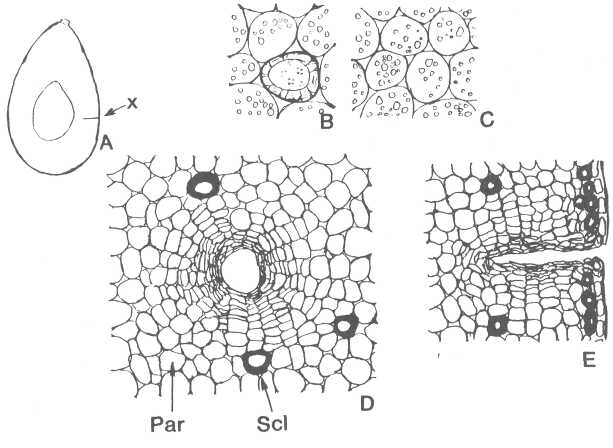
Figure 1. A - Diagrammatic longitudinal section through middle
of 'Hass' avocado fruit indicating a single puncture point (x). B - Transverse
section through a sclereid and surrounding
parenchyma. C - Section through pericarp parenchyma
with oil droplets. D - Transverse section near bottom of puncture wound showing
meristem together with sclereids
(SCL) and parenchyma (PAR). E - Longitudinal section through puncture wound
showing meristem and sclereids
near bottom of wound.
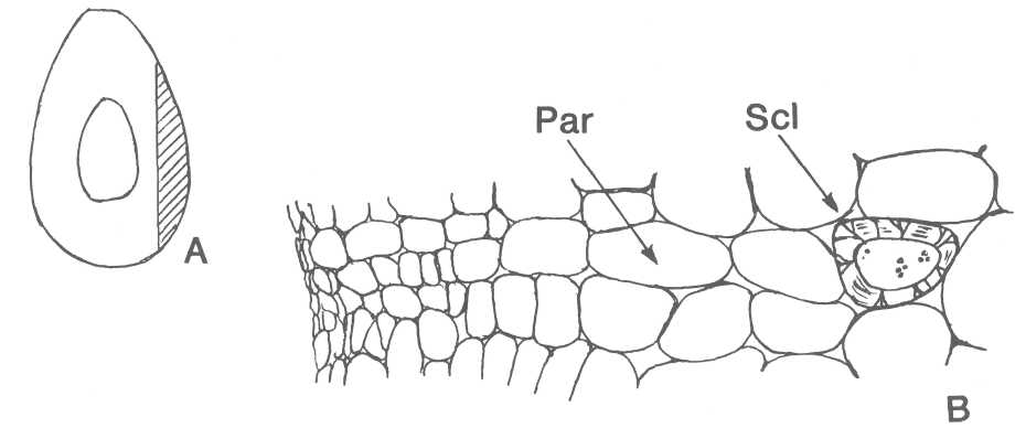
Figure 2. A - Diagrammatic longitudinal section of avocado
fruit along slit by a knife. B - Transverse section of tissue along the slit
showing meristem, parenchyma (PAR) and sclereid (SCL) at a distance from the injury.