Proc. of
Ultrastructure and Cytology of the
J.G. Luza and
CEPOC (Center for
Abstract. Basic postharvest work on 'Fuerte' avocado fruit cell wall anatomy and ultrastructure was carried out from fruit harvest to postclimacteric stage. Mesocarp samples were analyzed with light microscopy, scanning electron microscopy and transmission electron microscopy. Irregularity of the cell wall increased, microfibrils became disorganized, and some dissolution of the middle lamella was observed during ripening. However, the intercellular matrix remains almost unchanged without significant separation between the primary cell walls, as occurs in other fruits. This feature gives special consideration for avocado postharvest handling.
Mature avocado fruit start the ripening process following removal or abscission from the tree (Dallman et al., 1988). A characteristic of the ripening process, common to most fruits, is an increase in the activity of the cell wall degrading enzymes responsible for fruit softening. Dissolution of the ordered arrangement of cell wall and middle lamella polysaccha-rides occurs (Dick and Labavitch, 1989). Periodic Acid Schiff's Reagent (PAS) has long been used for polysaccharides as has Calcofluor for staining cellulose (Hughes and McCully,1975). These methodologies in combination with transmission electron microscopy (TEM) and scanning electron microscopy (SEM) studies have the ability to extend previous cytological information and open new avenues for additional research. For a better understanding of gas diffusion and controlled atmosphere applications in postharvest handling, it is necessary to solve questions concerning changes in mesocarp cells during ripening. The present study reports the cytological and structural cell wall changes during ripening in avocado fruit.
Materials and Methods
Sixty recently harvested, 'Fuerte1 avocado fruits were used. They were sorted into two groups: the first group was analyzed the day after harvest, the remainder analyzed at a post-climacteric stage, using both ripe and overripe fruit. Softening was determined with Mangness-Taylor pressure tester using a 7.9 mm plunger.
Samples for light microscopy were fixed in 4% glutaraldehyde, 0.2 M dipotassium
phosphate, 0.1M citric acid monohydrate, and 4% sucrose at pH 7.0. Dehydrated
samples were embedded in glycol methacrylate resin (DuPont-Sorvall;
SEM samples were submerged in freon for 1 minute and transferred to liquid nitrogen. Frozen samples were broken before the critical-point drying and were gold coated. TEM fixation was the same as LM, then washed, and post-fixed in 2% osmium tetroxide in buffer. The samples were embedded in Spurr's plastic (adapted from Dawes, 1979). Gold-colored sections were mounted on 300 mesh copper slim-bar grids (Ted Pella Inc.). The sections were stained in uranyl acetate and lead citrate solutions. Photographs and observations were made on a JEOL 100S TEM at 80kV.
Results
Stains used in this study revealed cellular events occurring during ripening of avocado mesocarp cell walls. Each stain gave different, but complementary information. In samples stained with calcofluor, cellulose walls fluoresced intensely while cytoplasmic components were completely unstained; calcofluor was useful for showing differences in cell wall thickness, cell division, and especially cell-to-cell "separation1. PAS-hematoxylin was a high contrast stain combination for revealing general structure and was specific for carbohydrate polymers. Flesh firmness of the recently harvested fruit measured 15.9 kg while the postclimacteric fruit measured 0.9 kg.
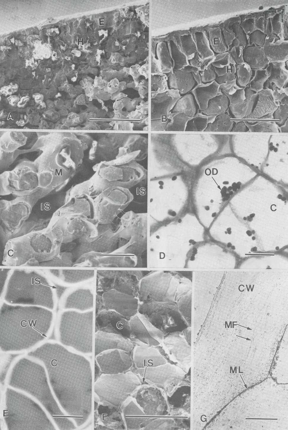
Anatomical analysis of fully mature fruit Only the exocarp and hypodermis show compact tissue (Fig. 1, A and B).The mesocarp has abundant oil droplets in the cytoplasm and oil sacs in scattered idioblasts. All of the tissue showed a strong positive reaction to PAS (Fig. 1, D), calcofluor staining is seen as bright, fluorescent rings, showing cell division after harvest (Fig. 1, E). LM shows a mesocarp formed by isodiametric cells, with tightly fitting cellulosic walls in adjacent cells. However, SEM shows a mesocarp with large intercellular spaces in relation to cell size (Fig. 1, C and F). The cells contained an intact plasmalemma tightly appressed to the walls and tonoplasts and nuclei bound by intact membranes. TEM analysis revealed cell walls of uneven thickness and composed of densely packed fibrils in an electron-translucent matrix. The middle lamella was visible as a more electron dense region between the primary walls (Fig. 1, G).
Anatomical changes in post-climacteric mesocarp The
plasma membrane draws away from the wall, cell wall unevenness increases, and
becomes more contorted following ripening. Cell wall thickening (swelling) becomes very irregular (uneven), a
characteristic found throughout the mesocarp,
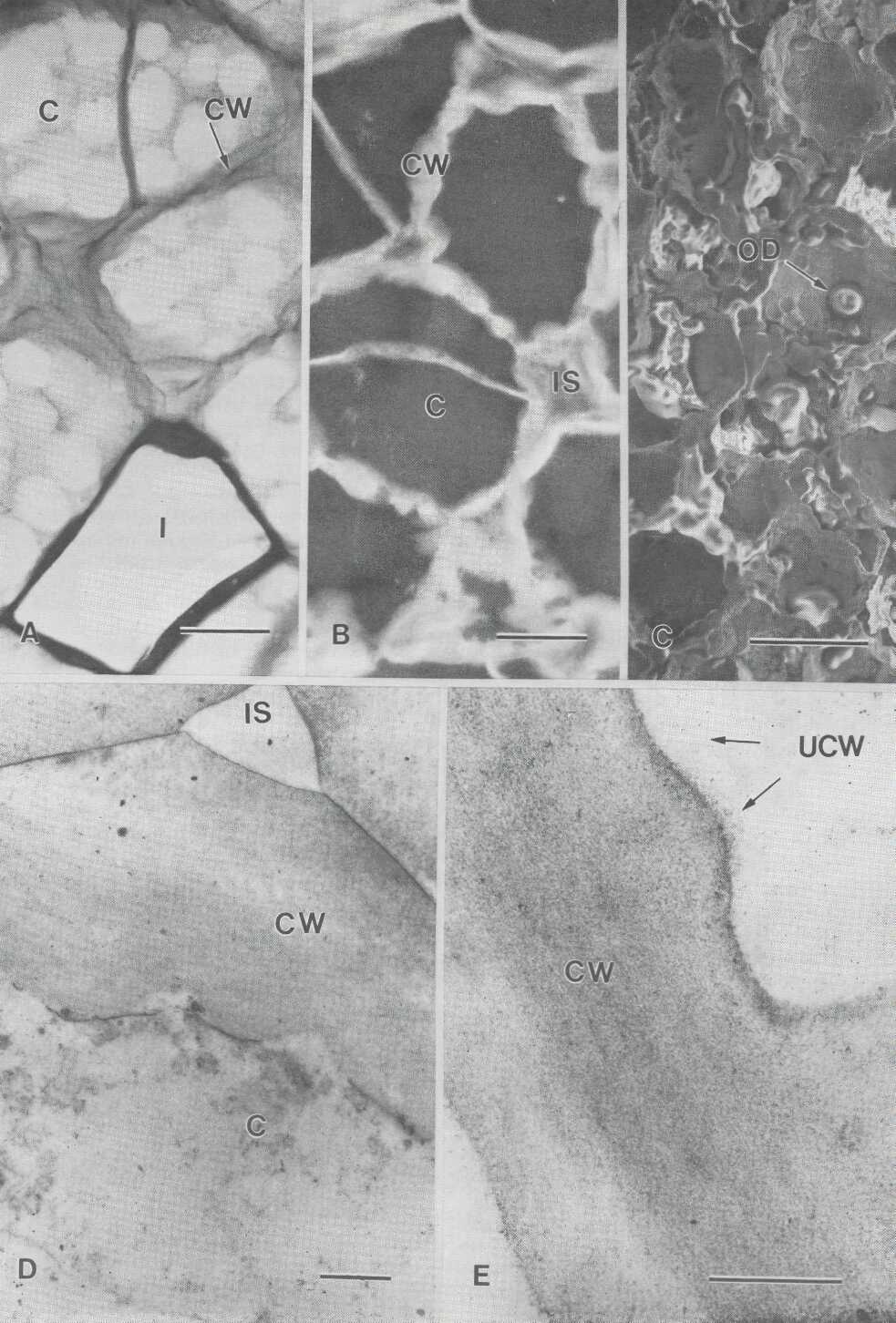
sometimes twice or more the normal thickness (Fig. 2, A and B). There is an
increase in both PAS and calcofluor staining as the
area of the cell wall increases. SEM shows both plasmalemma
and cytoplasm disruption. Oil is present as large drops over the inner cell
wall surface (Fig. 2, C). TEM reveals partial dissolution of the middle lamella
but the space between the primary cell walls does not increase. Microfibrils are no longer present and cell wall unevenness
is clearly present (Fig. 2, D and E).
Discussion
Plasmolysis and bursting during fixation is one of the main concerns in fruit cytological studies (Burns and Achor, 1989). Mature, ripe fruit mesocarp is very susceptible to disruption because of the delicate nature of the tissue itself, of which the main component are cells with large vacuoles and oil drops. A general analysis of our staining results tells us that the presence of polysaccharides and cellulose increased in a differential pattern during ripening. With PAS, a stain for insoluble polysaccharides, periodic acid oxidizes 1,2-glycol groups to formaldehyde groups (Jensen, 1962) which combined with Schiff's reagent, gives a colored product which is much more intense in postclimacteric fruits. Jewell and Saxton (1970) stated that it was the presence of arabinose and galactose in association with uronic substances in the middle lamella which were PAS positive. On the other hand, calcofluor staining patterns suggest the presence of cellulose in only primary cell walls. Structural changes in avocado cell walls became apparent during early softening stages due to some dissolution of the middle lamella and disintegration of cell wall fibrillar material. This is a well known alteration in the ripening process of avocado (Pesis et al., 1978), apple and pear (Ben-Arie et al., 1979).
Traditionally, avocado mesocarp
cells have been described as large polyhedral, isodiametric
cells, where all cell faces increase in area approximately evenly (Scott et
al., 1963, Platt-Aloia and Thomson, 1981).
However, SEM microphotographs show short cylindrical cells, narrow at the
contact points, and which have an intercellular
matrix proportional to cell size. Intercellular spaces did not increase during
ripening as is common in other fruit species. This feature in avocado fruit is
important to the understanding of gas exchange and gas diffusion during postharvest handling where pulp resistance plays a
fundamental role. Gas diffusion resistance of 'Gwen' avocado declines during
the postclimacteric phase after its rise during the
climacteric (Rodriguez et al., 1989). Shape and size of the cell and
intercellular network are specific for each cultivar. The cell surface exposed to
gas diffusion will depend on its surface-volume ratio and cell wall thickness.
The arrangement of internal cells will determine the percentage of
intercellular spaces (
Literature Cited
Ben-Arie, R., N. Kislev, and C. Frenkel. 1979. Ultrastructural changes in the cell walls of ripening apple and pear fruit. Plant Physiol. 64:197-202.
Burns, J.K. and D.S. Achor. 1989. Cell wall changes in juice vesicles associated with "section drying" in stored late-harvested grapefruit. Amer. Soc. Hort. Sci. 114:283-287.
Dallman, T.F.,
Dawes, C. J. 1979. Biological Techniques for Transmission and Scanning Electron Microscopy. Ladd Research Industries Inc. 303 pp.
Dick, A.J. and J. M. Labavitch. 1989. Cell wall metabolism in ripening fruit: IV.
Characterization of the pectic polysaccharides solubilized during softening of '
van Duijn, P. 1961. Acrolein-Schiff, a new staining method for protein. J. Histo. and Cyto. 9:234-241.
Hughes, J. and M.E. McCully. 1975. The use of an optical brightener in the study of plant structure. Stain Technol. 50:319-329.
Jensen,
W.A. 1962. Botanical
Histochemistry.
Jewell, G., and C. Saxton. 1970. The ultrastructural demonstration of compounds containing 1,2-glycol groups in plant cell walls. Histochem. J. 2: 17.
Labavitch,
J. M. 1988. Cell wall metabolism in fruit ripening.
Proceedings of the symposium on the physiology of fruit drop ripening, storage
and postharvest. Processing of
fruits, October 3-4,
Luza, J.G., R. van Gorsel, V.S. Polito, and A.A. Kader. 1991. Chilling injury in fruits of three peach cultivars: A cytochemical and ultrastructural cell wall study. J. Amer. Soc. Hort. Sci. (submitted).
O'Brien, T.P. and M.E. McCully. 1981. The Study of Plant Structure: Principles and
Selected Methods. Termarcarphi Pty. Ltd.,
Pesis, E., Y. Fuchs, and G. Zauberman. 1978. Cellulase and softening in avocado. Plant Physiol. 61: 416-419.
Platt-Aloia, K.A. and W. W. Thomson. 1981. Ultrastructure of the mesocarp of mature avocado fruit and changes associated with ripening. Ann. Bot. 48:451-465.
Rodriguez, L., D. Zagory, and A.A. Kader. 1989. Relation between gas diffusion resistance and fruit ripening in fruits. Proc. Fifth Int. Com. p. 2:1-9.
Scott, F., B. Bystrom, and
E. Bowler. 1963. Persea

Fig. 1. Mature tissue from recently harvested 'Fuerte' avocado fruit. A-C: exocarp
and mesocarp SEM microphotographs from a fully mature
fruit at harvest; A: bar= 28.8 mm. B: bar = 27.7 mm. C: bar = 18.9 mm. D-E: mesocarp with tightly fitting cellulosic
walls in adjacent cells, cell division is evident, bar = 17.3 mm.;D: PAS stained, E: calcofluor stained. F-G: EM microphotographs from mesocarp showing unevenness of the primary cell wall; F:
SEM, bar = 36.3 mm., G: TEM, bar = 18.3 mm., cell wall
shows packed fibrils, and middle lamella visible as a more dense region. C =
cytoplasm; CW = cell wall; E = exocarp; H =
hypodermis; IS = intercellular space; M = mesocarp;
ML = middle lamella; MF = microfibrils; OD = oil
droplets.

Fig. 2. (facing page) Observation from postclimacteric mesocarp of 'Fuerte' avocado fruit. A-B: cell wall thickening and unevenness is evident, bar = 17.3 mm., A: PAS stained. B: calcofluor stained. C-E: EM micrographs showing plasmalemma and cytoplasm disruption, no increase in the intercellular matrix, and microfibrils are no longer present. C: SEM, bar =22.7 mm., D: TEM, bar = 13.6 mm., E: TEM, bar = 24.6 mm. C = cytoplasm; CW= cell wall; I = idioblast; IS = intercellular space; OD = oil drop; UCW = uneven cell wall.